 Reproduced, with permission, from:
Holman, C. D. J., and B. K. Armstrong. 1984. Pigmentary traits, ethnic
origin, benign nevi, and family history as risk factors for cutaneous
malignant melanoma. Journal of the National Cancer Institute 72 (2):
257-66.
Reproduced, with permission, from:
Holman, C. D. J., and B. K. Armstrong. 1984. Pigmentary traits, ethnic
origin, benign nevi, and family history as risk factors for cutaneous
malignant melanoma. Journal of the National Cancer Institute 72 (2):
257-66.
C. D'Arcy J. Holman[3] and Bruce K. Armstrong[3, 4, 5]
ABSTRACT--The roles of constitutional factors and benign nevi in causation of malignant melanoma were examined in a case-control study of 511 patients and 511 matched controls in Western Australia. The strongest risk factor was the number of palpable benign nevi on a subject's arms. Compared to the risk of melanomas for persons having no palpable nevi on the arms, the relative risk of melanoma was 2.0 for persons with 1-4 nevi, 4.0 for persons with 5-9 nevi, and 11.3 for persons with 10 or more nevi (p<.0001). Of the several pigmentary traits known to have associations with melanoma, inability to tan was the most important. Susceptibility to sunburn and hair color also had significant effects that were independent of tanning ability; however, after these traits were controlled, measured skin color and eye color had no additional effects. A reduced risk of melanoma was observed in persons having two or more Southern European grandparents [odds ratio (OR)=0.39; P=.025]. Persons of Celtic origin did not have a significantly increased risk (IR=1.18). Possession of one or more affected blood relatives was related to an increased risk of melanoma (OR=2.69; p<.0001). The effects of pigmentary traits, benign nevi, ethnic origin, and family history as risk factors were largely independent of one another.--JNCI 1984; 72:257-266.
Several constitutional characteristics are widely accepted as risk factors for malignant melanoma of the skin. Melanoma is three to four times more common in lightly pigmented than heavily pigmented races (1). This proportion has been observed even when different races resided in the same geographic area and were served by the same cancer registry (2). Risk of melanoma within light-skinned (European) populations is also influenced by pigmentary characteristics and ethnic origin. The disease has been associated with fair skin, red or blond hair, blue or green eyes, and Celtic ancestry in case-control studies in Australia, the United States, and Great Britain (3-8). An association with self-reported tendency to burn in the sun has been less consistent (2, 5, 8, 9). In two studies, however, melanoma patients have shown a reduced minimal erythemal dose and an increased tendency to develop prolonged erythema in response to UV radiation (10, 11).
A role for heredity in determining melanoma risk is suggested by familial concurrence of the disease (12). An important, but not invariable precursor of familial melanoma is the dysplastic nevus syndrome, characterized by multiple atypical moles that continue to appear well into adulthood and occur most frequently on the trunk (13). In predominantly nonfamilial cases, longstanding pigmented lesions at the site of melanoma development have been described by some two-thirds of patients (14), and remnants of benign nevi have been identified histologically in approximately one-quarter of the patients (15).
There are, however, at least two deficiencies in the evidence that associates malignant melanoma with constitutional factors and benign nevi. First, the relationships have not been quantified. For example, measures of effect were not reported in any of the eight case-control studies that examined the associations of melanoma with pigmentary characteristics and ethnic origin (3-9, 16). The relationships of melanoma with family history and benign nevi have never been measured in a well-controlled analytic investigation. Second, the possibility of confounding between constitutional risk factors has not been considered. The established risk factors (e.g., skin, hair, and eye color) are highly correlated, and it is possible that some have apparent influence only by virtue of an association with a more fundamental factor. In addition to making good these deficiencies, there is a need to examine the role of constitutional factors and benign nevi in the occurrence of different morphologic subtypes of melanoma.
This paper aims to quantify the relative and independent contributions of pigmentary traits, ethnic origin, family history, and benign nevi to the occurrence of cutaneous malignant melanoma in a Caucasian population. It is the first substantial report of a study in which 511 patients with melanoma incident in Western Australia in 1980-81 and 511 population-based controls were interviewed and examined with respect to a wide range of constitutional and environmental factors.
Cases.---All cases of histologically diagnosed preinvasive (i.e., level I) and invasive melanoma, possible melanoma, and Spitz nevus occurring in Western Australia for the first time in 1980 or 1981 were identified from histopathology reports issued by public and private medical laboratories. Diagnoses taken as possibly indicative of preinvasive melanoma were HMF or SSM level I, in situ melanoma, premalignant or precancerous melanosis, and atypical melanocytic hyperplasia. A total of 820 melanomas arising in 803 patients were ascertained, a further 9 patients had lesions described as "possible melanomas," and 14 had Spitz nevi.
Histopathologic review was undertaken by a panel of 6 pathologists in 766 of the 820 preinvasive and invasive melanomas and in all cases of possible melanoma and Spitz nevus. Failure to review sections was due either to patients presenting with cryptogenic metastases (i.e.. melanoma metastases without an identifiable primary site, 15 cases) or to unavailability of slides (39 cases). At least 3 of the 6 pathologists examined sections from each tumor under blind study conditions. Among the features recorded were confirmation or rejection of a diagnosis of melanoma, histogenetic type according to the McGovern classification (17), level of invasion (18), and tumor thickness (19). Final details were recorded on the basis of majority decisions of the reviewing pathologists.
Diagnosis of melanoma was rejected by the panel in 10 of the 766 lesions initially diagnosed as melanomas and was accepted in 4 of the 9 possible cases and in 1 of the 14 Spitz nevi. Thus after the review process was complete, including acceptance of the original diagnosis in the 39 primary cases unavailable for review, the study series consisted of 815 melanomas arising in 798 patients.
All patients diagnosed in the period from January 1, 1980, to November 5, 1981, and aged less than 80 years were considered eligible for interview. Interviews were not attempted, however, in some remote areas of the State (containing about 3% of population) nor in those diagnosed after June 26, 1981, in some rural areas previously covered. Altogether 670 patients were eligible for interview.
Permission to approach each patient was sought from the attending surgeon and general practitioner. Permission was granted in 582 (87%) cases. Some patients were excluded by doctors because of mental retardation or dementia (6 cases) or because of death (11 cases).
Patients to be interviewed were contacted initially by mail, asked to participate in a study of "Environment, Lifestyle and Health," and given an appointment for the interview. They were telephoned on the day before the interview was scheduled to confirm the appointment. Those who could not be contacted by telephone were considered untraceable after three unsuccessful home visits by an interviewer, at least two of which were in the evening or on a weekend. Fifty-four patients refused to participate in the study, 15 were untraceable or known to have migrated from Western Australia, and 2 in rural areas could not be contacted before the end of the study. Thus of the 582 cases who could have been interviewed, 511 (88%) were interviewed. The response rate in cases actually approached for interview was 90% (511/565).
These 511 cases consisted of 233 males and 278 females whose ages ranged from 10 to 79 years. They displayed a typical Caucasian primary site distribution of melanoma with lesions mainly on the trunk (45%) in males and lower limb (39%) in females. In 29% of cases the melanoma was classified as preinvasive; a further 34% were thin melanomas (i.e., tumor thickness <0.76 mm). With respect to histogenetic type, 17% were HMF, 53% were SSM, 17% were UCM, and 10% were NM; in 14 cases (3%) the type was not known because sections were not reviewed by the pathology panel. The delay between diagnosis and interview of cases ranged from 31 to 443 days, with a mean of 118 days.
Controls.---For each case who was 18 or more years of age, a control of the same sex, born within the same 5-year period and resident in the same electoral subdivision, was selected from the Australian Commonwealth Electoral Roll by use of a table of random numbers. For 13 patients it proved difficult to find controls born within the same 5-year periods; for them, controls were selected as the nearest in age of 240 randomly selected individuals who were otherwise matched on sex and electoral subdivision. The age differences between cases and controls in these instances were either 5 years (10 pairs) or 6 years (3 pairs). Ten patients were aged less than 18 years at the time of control selection. For each of these patients, a control of the same sex and age was randomly selected from the student roll of a public school servicing the area of residence of the patient. School students were approached only with the consent of their parents pr guardians. Matching on age and sex was undertaken with the aim of controlling confounding on these variables. Matching on electoral subdivision or school area was performed for ease of study administration and to ensure that the members of each case-control pair could be interviewed by the same interviewer.
The methods used to establish contact with and to solicit the cooperation of potential controls were identical in every respect to those used for cases. As far as possible, interviewers and other persons who contacted subjects were unaware of their case-control status.
To obtain the final series of 511 matched controls it was necessary to approach 824 potential controls. Eighty were either untraceable or known to have migrated out of State, and 229 refused to participate. Two subjects were excluded because of dementia and 2 because they did not speak English and an interpreter was not available. Thus the response rate in controls who were approached and could have been interviewed was 69% (511/740). At the time of interview the mean age of controls was 46.68 (+/-16.73 SD) years and the mean age of cases, 46.86 (+/-16.72 SD) years. In all but 4 pairs, cases and matching controls were interviewed by the same interviewer.
Data collection.---Constitutional risk factors were assessed by interview, objective measurement of skin, hair, and eye color, and counting of palpable nevi on the arms. Subjects were also interviewed regarding history of the sun exposure, hormone use, diet, and several other factors; were asked to complete a 24-hour dietary record; underwent measurements of their weight, height, hairiness, and extent of actinic damage in their skin; and were asked to volunteer a venous blood sample for retinol and cholesterol assays. The results arising from these additional data will be the subject of future reports.
Interviews and measurements in the metropolitan area of Perth were conducted by 3 trained nurse interviewers. In rural areas interviews were conducted by 24 trained nurses who were otherwise engaged in community health work. At the beginning of the study all rural interviewers were taught interviewing techniques by an experienced interviewer who demonstrated the interview of 1 case-control pair. Most interviews (96%) were conducted in the permanent residence of the subject, and the remainder were conducted at the workplace of either the subject or the interviewer. In 99% of the interviews information was obtained directly from the case or control subject. Three patients and 6 controls who did not speak English were successfully interviewed with the aid of an interpreter. Information relating to the youngest patient and control (10 yr old) was obtained from their parents. Upon completion of the interview, subjects were rated according to the apparent reliability of the information they provided. Information on only 10 cases and 3 controls was rated as poor, mainly because these subjects had difficulty in providing detailed residential and occupational histories. Subjects with a poor reliability rating were not excluded from these analyses because constitutional variables were assessed either objectively or by comparatively simple questions.
Interview questions relevant to constitutional factors dealt with acute and chronic skin reactions to sunlight, ethnicity of grandparents, family history of melanoma or xeroderma pigmentosum, history of excision of benign or malignant moles, and treatment of cancers other than skin cancer. Questions that related to moles, melanomas, and other cancers were placed at the end of the interview to reduce rumination bias. The subjects' own assessments of their acute and chronic reactions to sun exposure were elicited by the following questions:
"If your skin was exposed to strong sunlight for the first time in summer for one hour, would you...
- Get a severe sunburn with blistering?
- Have a painful sunburn for a few days followed by peeling?
- Get mildly burnt followed by some degree of tanning?
- Go brown without any sunburn?"
"After repeated and prolonged exposure to sunlight would your skin become...
- Very brown and deeply tanned?
- Moderately tanned?
- Only mildly tanned due to a tendency to peel?
- Only freckled or no suntan at all?"
It was often necessary for interviewers to press the supposititious nature of these questions and to insist on a choice from one of the standard replies (offered on a display card) to avoid responses with a behavioral element such as "I never allow myself to get burnt."
The ethnic origins of the subject's 4 grandparents were recorded. The birthplace of each grandparent was also sought to avoid confusion between ethnicity and country of birth. Grandparents who had varied ethnic origins were assigned to the ethnic group that accounted for the greater part of their ancestry or, where two or more ethnic origins were represented equally, to the more lightly pigmented ethnic group. Of the 4,088 grandparents, 28% were known to have been born in Australia, but they could not be assigned to any specific ethnic group. These grandparents were recorded as "Australian" (a misnomer, as the only true ethnic Australian is the Australian Aborigine), although it is likely that most were English, Welsh, Scottish, or Irish.
Skin colors were measured at three sites---the dorsum of the left hand as a site of continuous sun exposure, the tip of the left shoulder as an intermittently exposed site, and the left upper inner arm as a site not usually exposed to sunlight. Skin color measurements were taken by metropolitan interviewers using goggles fitted with monochromatic filters (Wratten Gelatin Filter No. 90; Kodak Australasia, Perth) and a 32-step Munsell Neutral Value Scale (Munsell Color; Kollmorgen Corp., Baltimore, Md.) (20). In rural areas skin color was graded visually against a 10-step complexion chart designed by Scotto and Fears (21). Interviewers assessed eye color by inspection and recorded it as blue, brown, gray, green, or hazel. Natural hair color was graded visually against 23 samples of human hair selected from the JL International Colour Range (samples No. 1, 2, 3, 4, 6, 9, 11, 13, 17, 20, 22, 24, 24a, 25, 29, 31, 32, 33, 39, 54, 60, 103, and 107; Europa Haar Modelle, Fantastic Hair, London, England). Subjects who were bald, had gone gray, or had artificially colored hair were asked to select the hair sample that would have provided the best color match with their natural or original hair color.
Pigmented nevi were counted on the arms of each subject below the level of the axillae. Although a count of nevi on the whole body would have been desirable, the nonclinical setting of the interviews and difficulty in providing a "chaperone" prevented it. To avoid confusion with freckles, interviewers counted only nevi that were palpably raised above the surrounding skin. This procedure would have biased the count in favor of compound and intradermal nevi and against junctional nevi.
Methods of analysis.---The analysis followed the methods described by Breslow and Day (22) for matched case-control studies. Exposure OR (estimates of the incidence density ratio in exposed and unexposed individuals) were calculated by conditional maximum likelihood estimation. Continuous or multiple-level exposure data were usually grouped into 4 categories so that approximately one-quarter of the subjects were in each category. The significance of trends in OR was assessed by the chi-square formula for matched data given by Breslow and Day (22). The Y-scores used in trend tests were assigned as the midpoint values of the exposure intervals or, for risk factors without numeric values, by the tied rank method (23). Conditional logistic regression analysis of the simultaneous effects of two or more risk factors was performed by use of a FORTRAN program, "RISK" (24). For stepwise analyses, factors were incorporated into the model in a sequence determined by their contributions to goodness of fit as assessed by the log likelihood statistic (22).
Four controls reported a history of excision of a malignant mole. These controls and their corresponding cases were excluded from all analyses except the analysis of history of melanoma as a risk factor. After the analysis of all types of melanoma combined, most factors were examined for associations with each of the four histogenetic types of melanoma by the restriction of each analysis to cases with a particular histogenetic type and to their matched controls. More specific details of the methods of analysis of some risk factors are given below.
Pigmentary Traits and Reaction to Sunlight
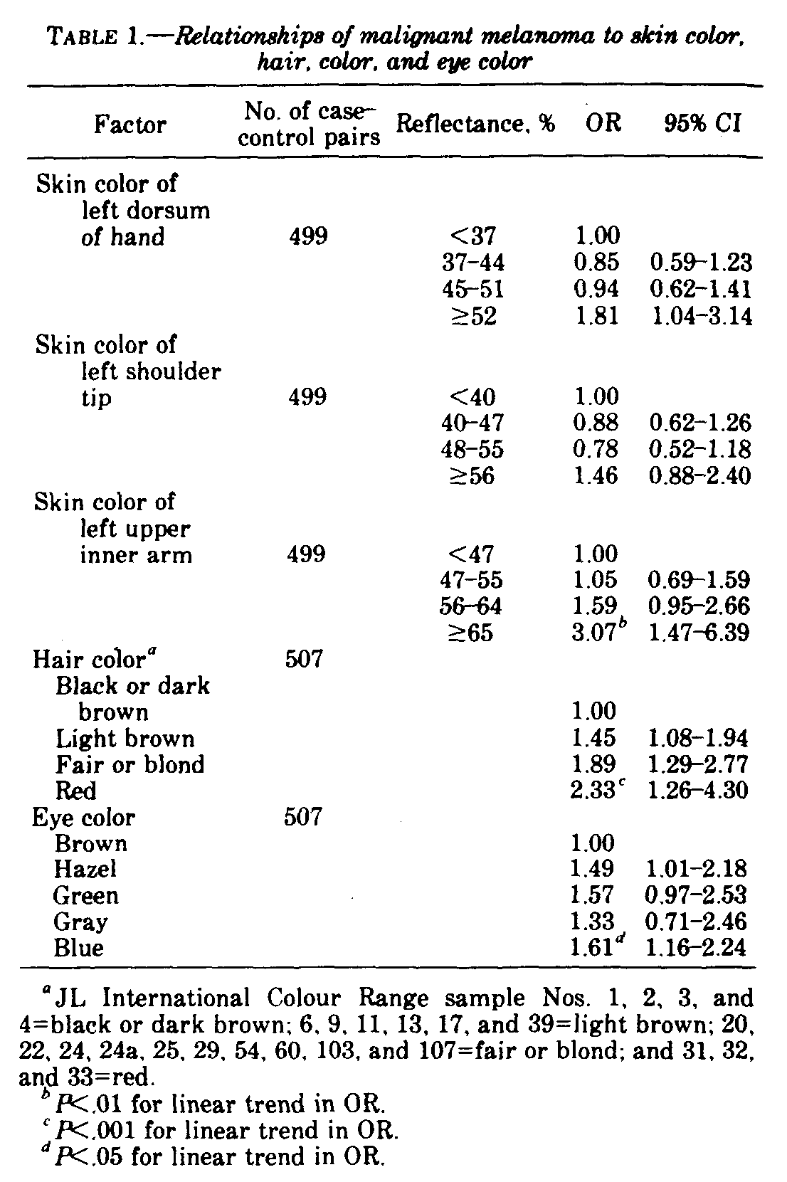
Results for skin color, hair color, and eye color as melanoma risk factors are shown in table 1. Skin color groups are given as ranges of reflectance values (20); a high reflectance is indicative of lightly pigmented skin. Skin colors measured in rural areas were converted to reflectance values for combined analysis, because it was thought that both methods of skin color measurement provided adequate discrimination for the purpose of categorizing subjects into 4 groups. Skin color tended to be darkest on the dorsum of the hand, lighter on the shoulder tip, and lightest on the upper inner arm; thus reflectance values were grouped differently for each site to maintain an even distribution of subjects across the groups.
No consistent relationship was seen between risk of melanoma and skin color of the dorsum of the hand or of the shoulder tip, although a tendency toward an increased risk in persons in the fairest skin color group was apparent in both instances (table 1). There was, however, a much stronger and statistically significant association with skin color of the upper inner arm: Persons in the fairest group experienced a threefold greater risk than those with dark pigmentation (reflectance, <47%). Significant results were observed also in relation to hair color and eye color. Persons with red hair were at more than a twofold greater risk than those with black or dark brown hair, whereas intermediate risk levels were seen in persons with light brown and fair or blond hair. Variation in risk with respect to eye color was less, although still significant at the 5% level. The highest OR of 1.61 was found in subjects with blue eyes, and the second highest was seen in those with green eyes.
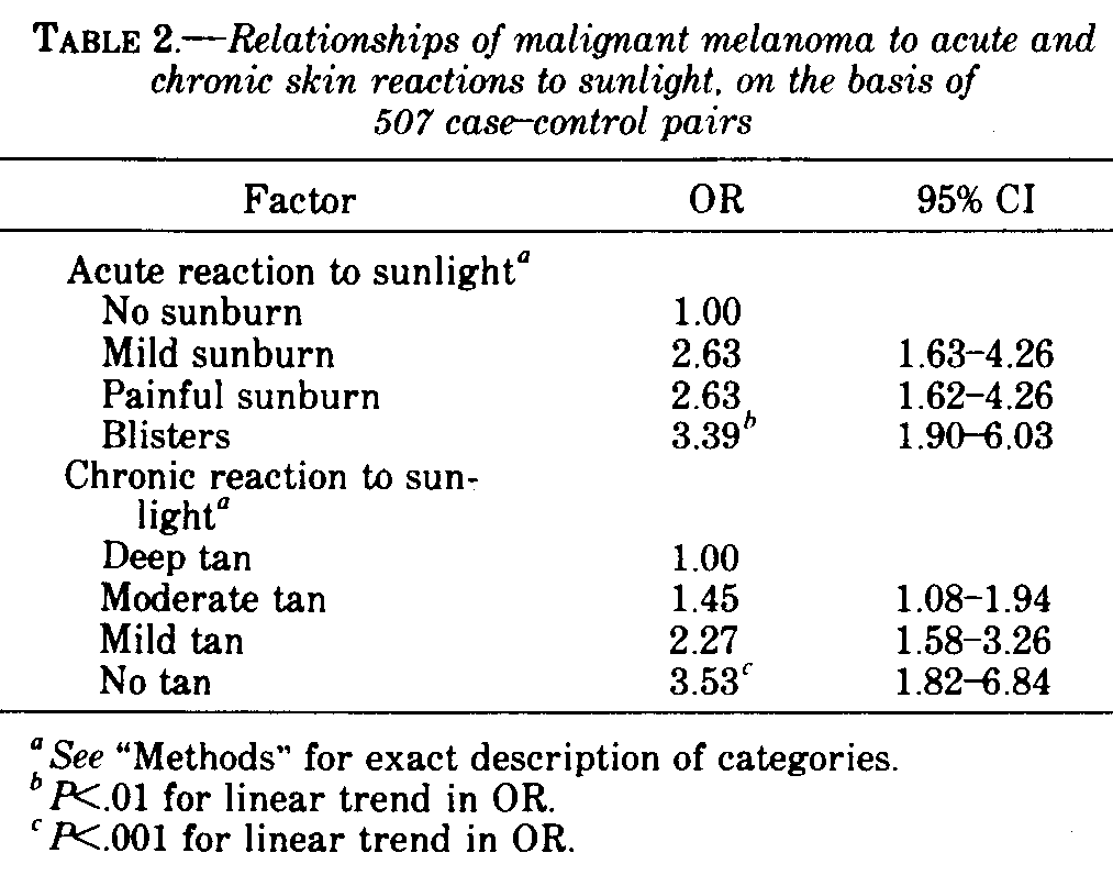
Table 2 gives results in relation to the subjects' assessments of their acute and chronic skin reactions to sunlight. Melanoma risk was strongly and significantly related to both factors, the highest OR occurring in persons whose acute reaction was to blister or chronic reaction was to freckle rather than to tan.
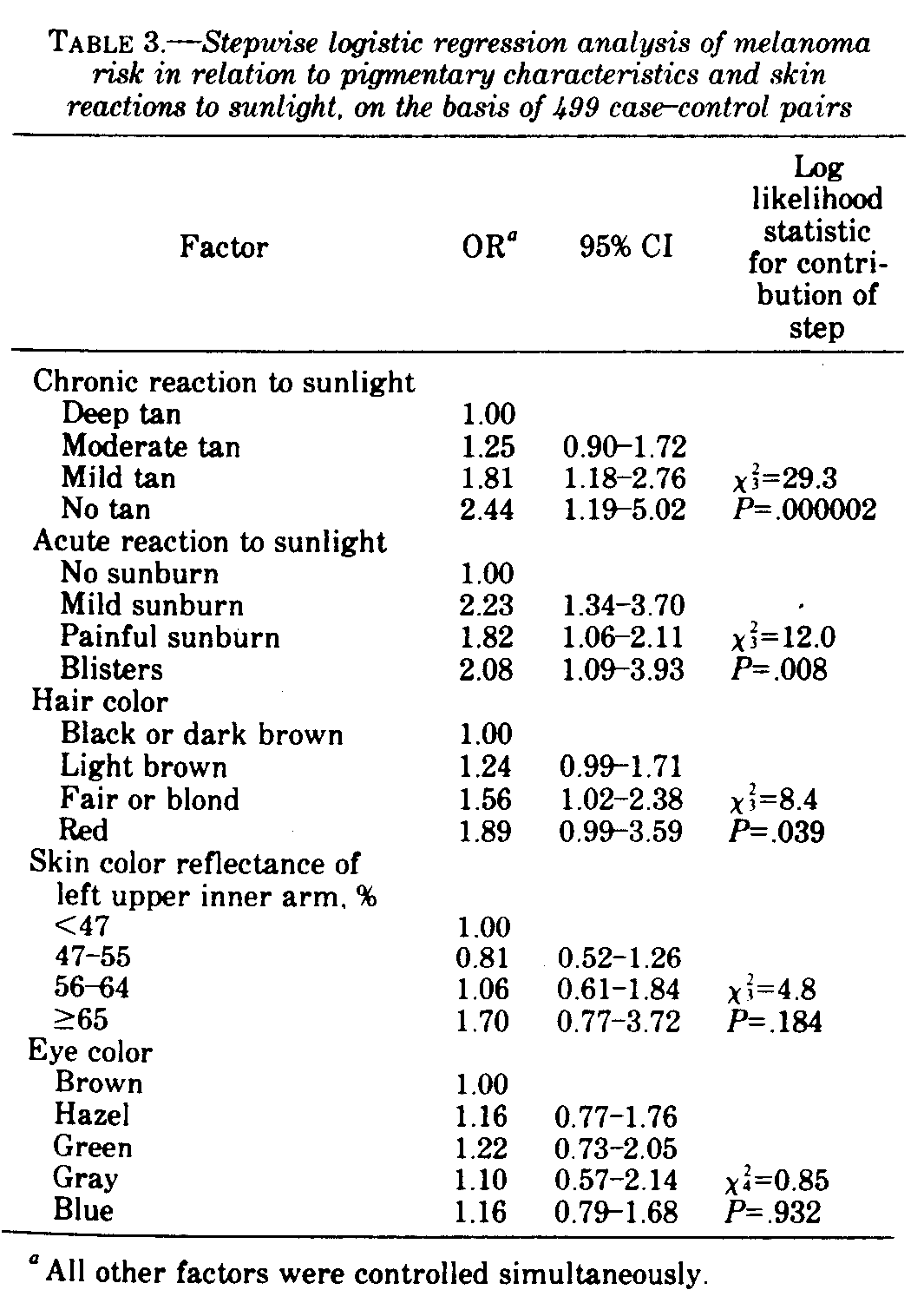
A stepwise logistic regression analysis was undertaken to identify which of the factors described above had independent effects. The results, summarized in table 3, identified chronic reaction to sunlight as the most important risk factor, followed by acute reaction to sunlight and hair color. After taking these three factors into account, skin color of the upper inner arm and eye color did not improve goodness of fit and were associated with much reduced estimates of effect compared with the corresponding crude OR given in table 1. It should not be inferred from table 3, however, that hair color is superior to skin color as a melanoma risk indicator. When skin color, hair color, and eye color were examined separately from acute and chronic reactions to sunlight, the greatest independent contribution to goodness of fit was made by skin color followed by hair color which also had a significant effect. It appears, therefore, that the effects of skin color were explained almost entirely by chronic and acute reaction to sunlight, whereas hair color persisted in having an independent effect. Further analyses also showed that the independent effect of acute reaction to sunlight resulted from a low risk in persons who never burnt, with little additional effect given by the severity of burn reaction in other subjects. This finding is shown in table 3 by the similarity and unordered pattern of OR associated with degree of burn reaction.
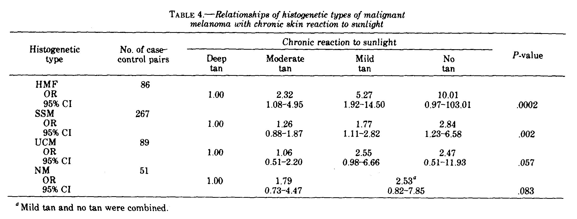
Relationships of each of the four histogenetic types of melanoma with chronic skin reaction to sunlight are shown in table 4. Risk of all four types increased with decreasing ability to tan, although the association was much stronger for HMF than for SSM, UCM, or NM. Each of the histogenetic types was also related to acute reaction to sunlight, hair color, skin color of the upper inner arm, and eye color, with the exception that HMF and UCM did not appear to have any relationship with eye color.
Ethnic Origin
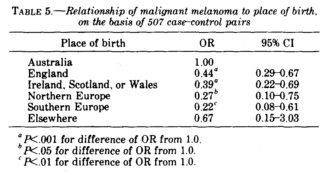
Ethnic origins of grandparents were categorized as Celtic (Irish, Scottish, or Welsh), English, Australian (probably Celtic or English), Southern European (Albanian, Bulgarian, Greek, Italian, Maltese, Portuguese, Spanish, or Yugoslav), Northern European (all other ethnic groups in Europe), African, or Asian. The analysis of ethnic origins was complicated by a confounding effect of age at arrival in Australia. Previous descriptive studies have shown that immigrants to Australia are at a substantially lower risk of melanoma than are native-born Australians (25, 26). This observation was evident also in this study (table 5); a reduced risk was observed in all persons born outside Australia, regardless of their country of birth. Further analysis, which will be described in detail elsewhere, showed that the main factor affecting level of risk in immigrants was age at arrival in Australia. Because subjects with mainly English or Celtic grandparents were more likely to have been born in Australia than those with other ethnic backgrounds, control of age at arrival in Australia was necessary in the analysis of ethnic origin. A further difficulty was presented by positive and negative correlations between ethnic groups themselves. For example, a subject with 2 Celtic grandparents had a reduced likelihood of also having 2 Southern European grandparents. Thus any effect observed with Celtic grandparents could be interpreted as an effect caused by lack of Southern European grandparents.
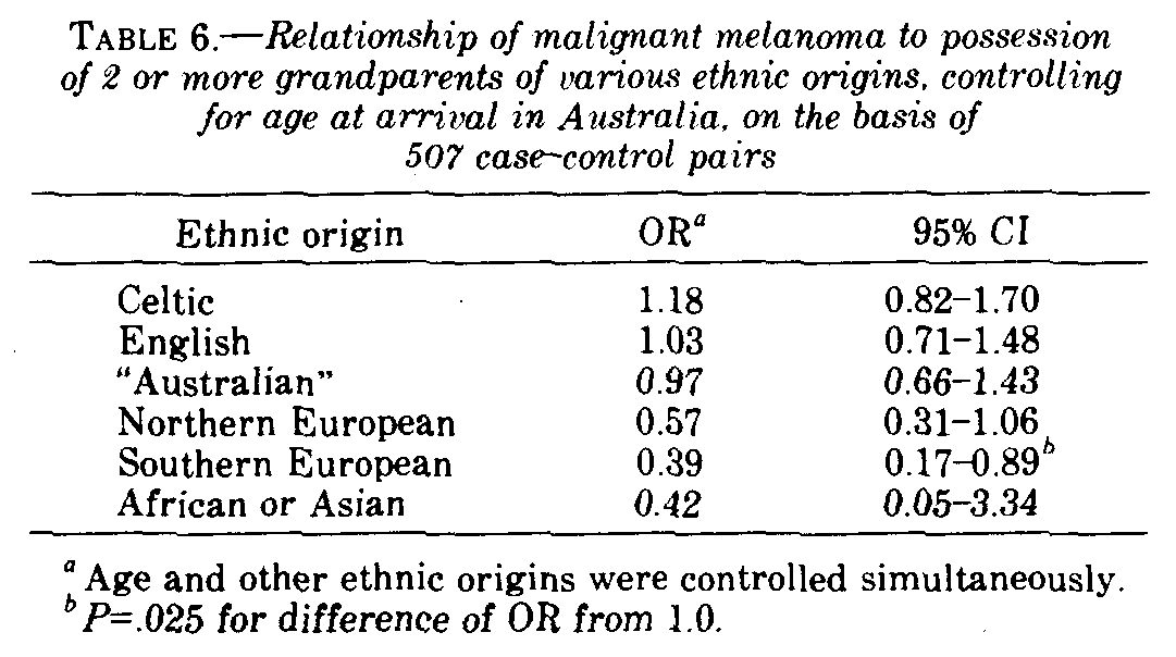
Results of a logistic regression analysis designed to separate the independent effects of having 2 or more grandparents belonging to various ethnic groups and to control for confounding by age at arrival in Australia are given in table 6. Possession of 2 or more Southern European grandparents was significantly protective from risk. Low OR were also seen with Northern European and African or Asian grandparents, although for those with African or Asian grandparents the 95% CI was wide. No significant independent effects were observed with possession of Celtic, English, or Australian grandparents. The crude OR associated with 2 or more Celtic grandparents was 1.32 (95% CI=0.98-1.77; P=.070), which fell to 1.26 (95% CI=0.94-1.59; P=.128) when age at arrival in Australia was taken into account and fell further to 1.18 (table 6) when the simultaneous effects of other grandparents were also controlled.
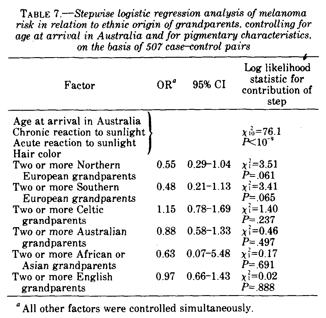
To see whether protective effects of Southern or Northern European grandparents could be explained by differences in chronic and acute reactions to sunlight and hair color, we performed a stepwise logistic regression analysis (table 7). Additional contributions to goodness of fit made by both Northern European and Southern European ethnicity, after pigmentary traits and age at arrival in Australia were taken into account, were not significant at the 5% level. Comparison of the OR in table 7 with those in table 6 shows that the strength of the protective effect of Southern European grandparents was reduced when confounding by pigmentary traits was controlled. This was not so for the effects of Northern European grandparents.
Benign Nevi
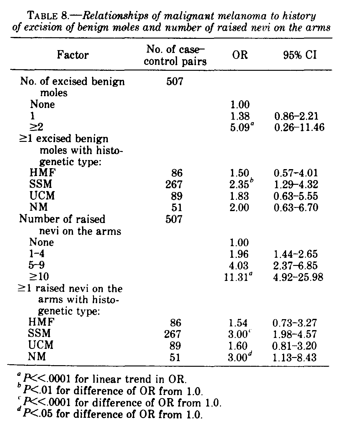
The associations of melanoma with number of palpable nevi on the arms and history of surgical excision of 1 or more benign moles are shown in Table 8. An excised mole was considered to have been a nevus only if the subject's doctor had said it was benign (given the many factors that might influence the decision to remove a mole, a history of mole excision must be regarded as only an approximate indicator of "mole-proneness"). Nonetheless, both the number of nevi on the arms and history of mole excision were strong risk factors for melanoma. The association with nevi on the arms was especially strong; persons with 10 or more nevi had an OR of 11.3 in comparison with those who had none. The association with nevi was strongest for SSM and weakest for HMF.
Family History of Melanoma
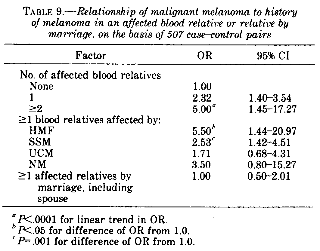
Findings in relation to family history are given in table 9. A positive history in a blood relative was reported by 15% of cases and 6% of controls. These numbers resulted solely from the interviews; medical documentation was not sought. A history of melanoma in a blood relative was a strong and statistically significant risk factor. When examined by histogenetic type, the association with a positive family history was strongest for HMF, intermediate for SSM and NM, and weakest for UCM. Although HMF is usually diagnosed at an older age than other histogenetic types, age could not have been a confounding factor in the analysis of family history because all cases were matched to controls of approximately the same age. Spouses with a history of melanoma were reported by 11 cases and 9 controls. There was no increase in risk with an affected spouse or other relative by marriage. The mean number of palpable nevi on the arms was no higher in those with a family history of melanoma (3.37) than in those without (3.31).
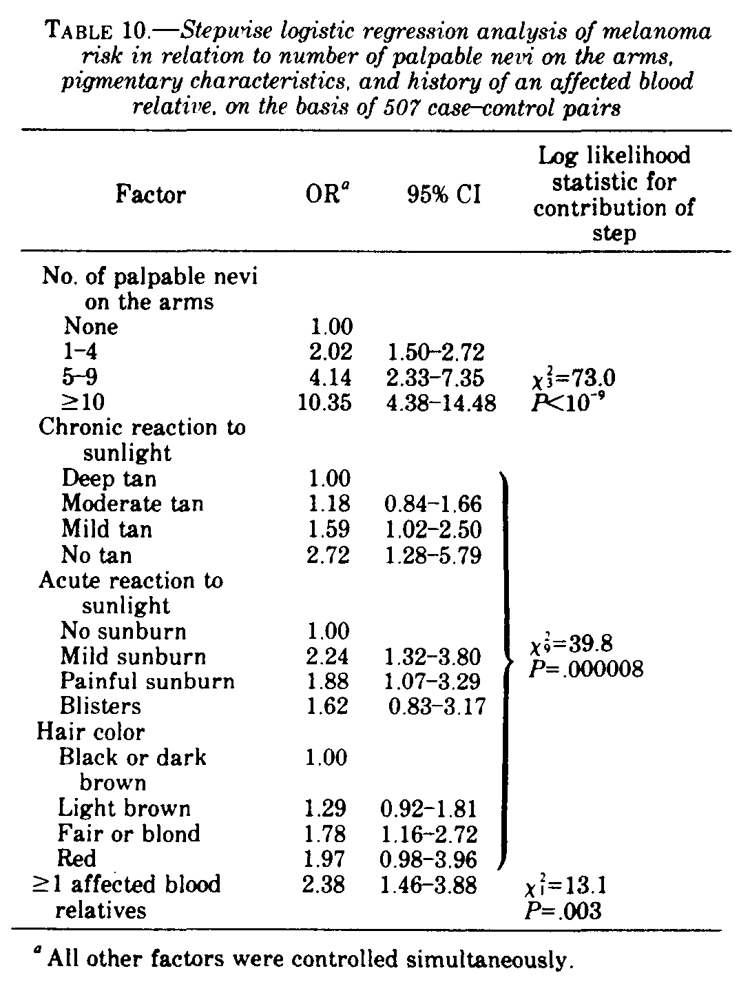
In table 10 the results of a stepwise logistic regression analysis show the independent effects and contributions to goodness of fit of number of raised nevi on the arms, pigmentary characteristics, and family history. All three sets of factors had significant independent effects on melanoma risk, the strongest influence being that of number of palpable nevi on the arms. In the comparison of the OR of 2.38 associated with family history in table 10 with the crude OR of 2.69 (95% CI=1.79-4.05; P=.000002), it was apparent that controlling for the effects of number of nevi and pigmentary traits made only a small difference in the observed risk level in persons with an affected blood relative.
In addition, inclusion of ethnic origin of grandparents and age at arrival in Australia in the logistic model had little effect on the OR estimates and their significance levels shown in table 10. Similarly, the parameter values for the various ethnic origins of grandparents were only slightly different from those shown in table 7.
Other Factors
Eighteen cases and 4 controls reported a history of a mole excised before 1980 that their doctor said was malignant (OR=4.50; 95% CI=1.44-15.69; P=.006). No statistically significant relationship was seen with history of noncutaneous cancer (OR=1.50; 95% CI=0.643.58; P=.424). History of nonmelanoma skin cancer will be dealt with elsewhere. Two melanoma patients claimed to have had a blood relative affected by xeroderma pigmentosum.
Possible Sources of Bias
This study may have been subject to several types of bias which one should keep in mind when interpreting the results. A selection bias may have been introduced, because only 69% of controls approached were interviewed, in contrast to 90% of approached cases. Since the effort made to obtain cooperation was the same in the 2 groups, other factors, such as less interest in health in the general population (potential controls) than in people who had recently undergone surgery (potential cases), must have intruded. Nonparticipation in survey research is well known to relate to socioeconomic status and life-style characteristics (such as alcohol and tobacco consumption) (27, 28) and presumably has its origin in the underlying personality, among other things. Whether personality is also related to the constitutional characteristics investigated here is unknown, but it seems unlikely.
Recall and rumination bias in the subjects and interviewer bias were minimized, as far as possible, by concealing the main purpose of the study from subjects and keeping interviewers blind to the case-control status of each subject. On occasions, the melanoma patients guessed that their involvement was consequent on recent treatment for melanoma, and the interviewers sometimes learned the status of the subject before or early in the interview. Thus opportunities for bias did arise. Their effects, however, particularly on semiobjective measurements (e.g., measurement of skin and hair color and nevus counts), should have been small.
Pigmentary Characteristics and Response to Sunlight
The increased risk of melanoma in persons with fair skin complexion, red or blond hair, and blue eyes observed in this study is similar to that found in previous studies (3-5, 8). The associations observed with poor tanning ability and tendency to severe sunburn are also in general agreement with the results of other studies (3, 8), although in these earlier studies acute and chronic reactions to sunlight were not distinguished. We made special efforts to ensure that assessments by subjects of their skin reactions were not influenced by sun-avoidance behavior. Failure to consider this problem may explain some of the inconsistencies previously reported in the relationship of melanoma to skin reaction to sunlight (3, 5, 8, 9).
That the strongest pigmentary risk factor was chronic skin reaction to sunlight has been observed also in nonmelanotic skin cancer. Vitaliano and Urbach (29, 30) found that ability to tan was a more important indicator of risk of basal and squamous cell carcinomas than skin complexion. A similar conclusion had been reached by Silverstone and Searle (31). These results suggest that the ability to tan quickly in response to sunlight is of prime importance in reducing risk of skin cancers and is more important than the base-line skin color.
That acute reaction to sunlight and hair color had significant effects, after chronic reaction to sunlight was taken into account, does not necessarily mean that they operate through different mechanisms. The categorization of ability to tan into 4 discrete groups imposes arbitrary constraints on a characteristic that probably varies continuously. It may be that cross-categorization by acute reaction to sunlight and hair color merely provides sharper definition of the skin's capacity to produce a protective tan.
The pigmentary characteristics discussed above influenced risk of each histogenetic type of melanoma as well as melanomas in general. In the comparison of histogenetic types, however, HMF was the most strongly associated with poor tanning ability and other characteristics typical of fair-skinned individuals. This finding is consistent with the assertion that the causal relationship of sunlight with HMF is more direct than with SSM, UCM, or NM (32).
Ethnic Origin
No significant association was observed between melanoma occurrence and Celtic origin. Failure to control for the effects of immigrant status and the simultaneous effects of other ethnic backgrounds may have contributed to the much stronger associations reported in previous studies (6-8). Even when these effects were ignored in this study, however, the increased risk to persons of Celtic origin was of only marginal strength and significance. Misclassification of a proportion of subjects substantially of Celtic origin to the Australian category may have artificially reduced the apparent effects of having grandparents described specifically as Irish, Scottish, or Welsh.
The finding of a reduced risk of melanoma in persons of Southern European origin is consistent with the low incidence rates reported from Spain and other Latin populations (33). It would seem reasonable to attribute this effect, in part at least, to the darker pigmentation of Southern Europeans compared with the pigmentation of other Caucasians. This view gains only partial support from the increase in relative risk observed in Southern Europeans when pigmentary characteristics were controlled. An alternative explanation is that persons of Southern European origin may differ from other Australians in their pattern of sun exposure or with respect to other risk factors perhaps related to culture or socioeconomic status.
Family History
A family history of melanoma was more common in this study than in other case series (12). The high incidence of melanoma in Western Australia may explain this difference (25). The difference between cases and controls, however, showed that a positive history in a blood relative was a significant risk factor. This effect of family history was, for the most part, independent of mole proneness and pigmentary traits. The absence of an increased risk of persons with an affected spouse or other relative by marriage argues against an explanation of familial aggregation by shared environment. It appears, therefore, that there may be specific genetic factors that predispose to the development of melanoma.
Benign Nevi
The number of raised pigmented nevi counted on the arms was the strongest of any other risk factors described here. This observation may mean that a substantial proportion of melanomas have their origin in benign nevi---a suggestion supported both by studies of clinical history and histopathology (14, 15). In this respect our count of palpable nevi on the arms may not have been highly correlated with the total of all nevi on the body with malignant potential; i.e., substantial misclassification may have been present. Thus the real association of melanoma with nevi may be stronger than is suggested by this study. An alternative or perhaps complementary possibility to the origin of melanoma in nevi is that nevi may be markers of a constitution or environmental background that is associated with increased risk of melanoma arising in apparently normal skin.
Whether nevi should be regarded as of mainly constitutional or environmental origin is moot at present. Certainly, the dysplastic nevus syndrome shows familial aggregation (13), but dysplastic nevi, which progress to melanoma, are also seen in the absence of a family history (34). Even in familial dysplastic nevus syndrome, it may be the tendency to progress to cancer rather than the presence of the nevi that is constitutionally determined. Clinically, however, apparently normal nevi are more prominent, perhaps more frequent, and more likely to appear on sites such as the buttocks in subjects with dysplastic nevi than in other subjects.
Our results showed SSM to be much more strongly associated with number of nevi than HMF; NM and UCM occupied intermediate positions. This finding is consistent with a lack of clinical and histologic evidence for the origin of HMF in benign nevi (35) and provides further evidence for the view that HMF differs in its etiology from other melanomas (36). We have suggested that some nevi contain "initiated" cells that may develop into SSM and sometimes into UCM and NM as a result of promotion by intermittent sunlight exposure (36). Thus further study of the interaction between nevi and sun exposure habits is indicated.
In view of the probable importance of nevi either as an early stage in the pathotgenesis of non-HMF melanomas or as indicators of increased risk, expenditure of more effort in the study of factors that cause benign nevi in humans seems desirable. There is a need for more population-based data on the prevalence, site distribution, and histologic features of nevi. Moreover, documentation of causal factors, perhaps by case-control or cohort studies, might lead to valuable insights into the prevention of melanoma. Our results should not be interpreted as evidence that prophylactic excision of asymptomatic nevi is justified. The lifetime risk of melanoma developing in any one nevus is quite small. (We estimate the risk in Australia as approximately 1 in 1,000.)
Basic Constitutional Factor
Multiple variable analysis did not identify any single variable from those variables discussed above that could account for the effects of all the others. Eye color and skin color appeared unimportant when hair color and acute and chronic reactions of the skin to sunlight were taken into account. These pigmentary characteristics, however, appeared to explain only a part, if any, of the associations with ethnic origin, family history of melanoma, and number of nevi. The last three factors, in their turn, appeared to be largely independent of one another. All of these factors, therefore, at least in this study, must be considered as potential confounders when environmental variables are considered.
1 Received Nay 23, 1983; accepted August 25, 1983.
4 Address reprint requests to Dr. Armstrong.
Abbreviations Used: CI=confidence interval; HMF+Hutchinson's melanotic freckly; NM=nodular melanoma; OR=odds ratio(s); SSM=superficial spreading melanoma; UCM=unclassifiable melanoma.
(1) Crombie, IK. Racial differences in melanoma incidence. Br J Cancer 1979: 40:185-193
(20) Gibson IM. Measurement of skin colour in vivo. J Soc Cosmet Chem 1971; 22:725-740.
(24) Thomas DC. Program "RISK". Montreal: McGill Univeristy, 1980.
(35) Mishima Y. Melanocytic and nevocytic malignant melanomas. Cancer 1967, 20:632-649.